+918042781261

DRSHREEPALJAINPEDIATRICCARDIOLOGIST https://www.bestpediatriccardiologist.com
Preview

This is your website preview.

Currently it only shows your basic business info. Start adding relevant business details such as description, images and products or services to gain your customers attention by using Boost 360 android app / iOS App / web portal.

919167383837

Treatments

Pediatric heart disease refers to any heart condition or abnormality that affects children, from birth through adolescence. These conditions can range from congenital (present at birth) to acquired heart diseases, and they may involve defects in the heart's structure, blood vessels, or the functioning of the heart itself. Early detection, diagnosis, and management are crucial for improving outcomes and quality of life for children with heart disease.Types of Pediatric Heart Disease:Congenital Heart Disease (CHD):Congenital heart defects are structural abnormalities in the heart that occur during fetal development. These are the most common type of heart disease in children.Common types of congenital heart disease include:Atrial Septal Defect (ASD): A hole in the wall between the two upper chambers of the heart (atria).Ventricular Septal Defect (VSD): A hole in the wall between the two lower chambers of the heart (ventricles).Tetralogy of Fallot: A combination of four heart defects, including a VSD, pulmonary stenosis, right ventricular hypertrophy, and an overriding aorta.Coarctation of the Aorta: A narrowing of the aorta, which can restrict blood flow to the body.Transposition of the Great Arteries (TGA): The positions of the aorta and pulmonary artery are reversed, leading to improper circulation of oxygenated and deoxygenated blood.Hypoplastic Left Heart Syndrome (HLHS): A condition where the left side of the heart is severely underdeveloped, requiring immediate intervention.Patent Ductus Arteriosus (PDA): A condition where the ductus arteriosus, a blood vessel that connects the pulmonary artery to the aorta, fails to close after birth.Single Ventricle Defects: These involve having only one functional ventricle (instead of two), which affects the heart's ability to pump blood effectively.Acquired Heart Disease: While congenital heart disease is the most common form, children can also develop heart disease due to infections, inflammation, or other factors. These acquired conditions include:Kawasaki Disease: An inflammatory condition that can cause swelling in the blood vessels, including the coronary arteries, and may lead to heart damage.Rheumatic Heart Disease: Resulting from rheumatic fever, a complication of untreated strep throat, this disease can cause damage to the heart valves.Myocarditis: An inflammation of the heart muscle, often caused by viral infections, which can weaken the heart's ability to pump blood effectively.Endocarditis: An infection of the inner lining of the heart, typically affecting the valves, and often caused by bacteria entering the bloodstream.Arrhythmias: Abnormal heart rhythms, which can sometimes be congenital or acquired due to infections, medications, or other conditions.Symptoms of Pediatric Heart Disease:The symptoms of heart disease in children can vary depending on the specific condition and its severity. Common symptoms include:Cyanosis: A bluish tint to the skin, lips, or nails, indicating low oxygen levels in the blood.Rapid or Labored Breathing: Difficulty breathing or an increased respiratory rate, especially during activity or feeding in infants.Fatigue: Children with heart disease may tire easily, even with minimal exertion.Poor Feeding or Weight Gain: Infants may have trouble feeding and may fail to gain weight as expected.Chest Pain: Though rare in young children, chest pain may occur, especially in older children or those with arrhythmias.Swelling: Swelling of the feet, ankles, or abdomen may occur in cases of heart failure.Heart Murmur: A heart murmur, which is an abnormal sound during a heartbeat, is often one of the first signs of a heart condition.Palpitations or Irregular Heartbeats: Some children may complain of a sensation of their heart racing or skipping beats.Diagnosis of Pediatric Heart Disease:Diagnosing heart disease in children typically involves a combination of the following methods:Physical Examination:The doctor will check for signs such as heart murmurs, cyanosis, abnormal pulse, or signs of fluid retention.Echocardiography:An ultrasound of the heart that allows the doctor to visualize the heart's structure and blood flow in real-time. It is the most common and reliable imaging technique for diagnosing congenital heart defects.Electrocardiogram (ECG):Measures the electrical activity of the heart and can help identify arrhythmias or abnormal heart rhythms.Chest X-ray:Provides an image of the heart and lungs, which can help in identifying enlarged heart chambers or other structural problems.Cardiac MRI or CT:These advanced imaging techniques can be used to assess the heart's anatomy, particularly in more complex cases or when other tests are inconclusive.Blood Tests:In cases of acquired heart disease (e.g., myocarditis or Kawasaki disease), blood tests can help diagnose the underlying cause, such as infections or inflammation.Pulse Oximetry:A non-invasive test that measures oxygen saturation levels in the blood and can help detect cyanosis or heart conditions affecting oxygen levels.Treatment for Pediatric Heart Disease:Treatment for pediatric heart disease depends on the specific condition, its severity, and the child's overall health. The treatment options may include:Medications:Diuretics: To reduce fluid buildup in cases of heart failure.ACE Inhibitors or Beta-blockers: To improve heart function and manage blood pressure.Antiarrhythmic Medications: To treat abnormal heart rhythms.Antibiotics: For bacterial infections like endocarditis or in the case of Kawasaki disease.Surgical Procedures:Many children with congenital heart defects will require surgery to correct the structural abnormalities. This may involve:Cardiac Surgery: To repair or replace heart valves, close septal defects (ASD or VSD), or correct defects like tetralogy of Fallot or coarctation of the aorta.Heart Transplant: For children with severe heart failure or end-stage heart disease that cannot be treated by other means.Catheter-Based Interventions:Some conditions, such as atrial septal defects or patent ductus arteriosus, can be treated with catheter-based procedures, where a catheter is inserted through the blood vessels to close the defect or widen a narrowed blood vessel.Cardiac Rehabilitation:Children recovering from surgery or with heart failure may benefit from a cardiac rehabilitation program, which includes supervised physical activity, education on lifestyle changes, and psychological support.Follow-Up Care:Regular check-ups with a pediatric cardiologist are essential for children with congenital heart disease, even if they have undergone successful surgery. Long-term follow-up helps in monitoring heart function, detecting complications, and providing ongoing care.Prognosis and Quality of Life:The prognosis for children with heart disease depends on the specific condition, its severity, and how early it is diagnosed and treated. Many children with congenital heart disease who receive early treatment can lead relatively normal lives, though they may require ongoing care and monitoring. In some cases, children with more complex heart conditions may face long-term health challenges, including the need for additional surgeries or treatments in adulthood.Children with heart disease are often able to participate in physical activities, but the level of intensity and the type of activity should be tailored to their individual condition and abilities, as advised by their cardiologist.Conclusion:Pediatric heart disease is a diverse and complex field that involves both congenital and acquired conditions affecting children’s hearts. Early detection and intervention are key to improving the outcomes for children with heart disease. With advancements in medical and surgical treatments, many children with heart disease now live longer, healthier lives. However, lifelong monitoring and care are often necessary to manage potential complications as these children grow older.

16, 1st Floor, Tender Hearts Clinic, Avighna IX, Opposite Ladoo Samrat Restaurant, Dr Baba Saheb Ambedkar Rd, Lal Baug, Mumbai, Maharashtra, India

Fetal echocardiography is a specialized ultrasound examination used to assess the heart of a fetus while it is still in the womb. This procedure allows doctors to detect heart defects or abnormalities in a fetus before birth, enabling early intervention and planning for any necessary treatment once the baby is born.Importance of Fetal Echocardiography:Fetal echocardiography is crucial because many congenital heart defects (CHDs) can be detected before birth, which helps in making early decisions about the management of the pregnancy and the care needed for the baby after delivery. Early detection of a CHD can improve the outcomes for the infant by allowing healthcare providers to prepare for the condition and plan any required interventions in advance.When Is Fetal Echocardiography Performed?Fetal echocardiography is generally done between 18 and 22 weeks of pregnancy, though it can sometimes be done earlier or later depending on the situation. The procedure might be recommended for a variety of reasons, including:Routine Screening:If the pregnancy is considered high-risk, fetal echocardiography can help in identifying potential heart defects early.Family History of Heart Disease:If there is a family history of congenital heart defects or genetic conditions related to the heart, the doctor may recommend fetal echocardiography.Abnormal Findings on Routine Ultrasound:If routine prenatal ultrasounds detect abnormalities that suggest a heart defect, fetal echocardiography can provide more detailed information.Maternal Health Conditions:Certain maternal conditions, such as diabetes, lupus, or phenylketonuria (PKU), can increase the risk of congenital heart defects in the fetus, so fetal echocardiography may be recommended.Increased Risk Based on Maternal Age:Women over the age of 35 are at a higher risk of having a baby with a congenital heart defect, and fetal echocardiography may be performed for closer evaluation.Chromosomal Abnormalities:If there are known chromosomal abnormalities, such as Down syndrome, that are associated with higher rates of heart defects, fetal echocardiography may be conducted to assess for heart issues.Abnormal Genetic Tests:If genetic tests suggest that the fetus may be at higher risk for congenital heart defects, fetal echocardiography may be used to investigate further.History of Heart Defects in Previous Pregnancies:If a previous child had a congenital heart defect, there may be a higher risk for the current pregnancy, warranting a fetal echocardiogram.How Is Fetal Echocardiography Performed?Fetal echocardiography is a non-invasive procedure and is similar to a routine ultrasound but with a focus on evaluating the fetal heart. Here is an overview of the process:Preparation:No special preparation is typically needed for the mother before the procedure. However, she may be asked to drink water before the exam to fill the bladder, as this may help provide better images.Procedure:The procedure is typically performed while the mother is lying on her back, similar to a standard ultrasound.A gel is applied to the abdomen, and a small device called a transducer is moved over the area. The transducer sends out sound waves and then picks up the echoes that bounce back, which are used to create images of the fetus.Detailed Imaging:Unlike a regular ultrasound, which is often used to assess fetal development in general, fetal echocardiography focuses specifically on the heart.It provides high-resolution images of the heart chambers, valves, blood vessels, and the overall function of the heart.The doctor will carefully assess the blood flow through the heart using color Doppler imaging, which can detect abnormal patterns of blood flow, often indicating a defect.Duration:The procedure can take anywhere from 30 minutes to an hour, depending on the complexity of the examination and the baby's position.What Can Fetal Echocardiography Detect?Fetal echocardiography can detect a wide range of congenital heart defects (CHDs), including:Atrial Septal Defect (ASD):A hole in the wall between the two atria (upper chambers) of the heart.Ventricular Septal Defect (VSD):A hole in the wall between the two ventricles (lower chambers) of the heart.Tetralogy of Fallot:A combination of four heart defects, including pulmonary stenosis, a ventricular septal defect, right ventricular hypertrophy, and overriding aorta.Coarctation of the Aorta:A narrowing of the aorta, which can obstruct blood flow.Transposition of the Great Arteries:The aorta and pulmonary artery are switched, causing blood to circulate incorrectly through the body and lungs.Hypoplastic Left Heart Syndrome:Underdevelopment of the left side of the heart, leading to severe heart function problems.Aortic Valve Stenosis:Narrowing of the aortic valve, which restricts blood flow from the heart to the body.Pulmonary Valve Stenosis:Narrowing of the pulmonary valve, which impairs blood flow to the lungs.Truncus Arteriosus:A single large artery arises from the heart, rather than the two normal arteries, leading to oxygenated and deoxygenated blood mixing.Endocardial Cushion Defects:Abnormal development of the inner lining of the heart and heart valves, leading to holes between the heart chambers and valve malformations.Single Ventricle Heart Defects:A condition in which only one of the heart's ventricles is developed, often requiring multiple surgeries for the child to survive.Why Is Fetal Echocardiography Important?Early Diagnosis:Detecting heart defects before birth allows doctors to plan for the delivery of a baby with a congenital heart condition, enabling early intervention and improved outcomes.Preparation for Birth:In some cases, specialized care may be needed immediately after birth, such as surgery or medication to stabilize the baby’s heart function. Early detection ensures that the appropriate medical team and resources are available.Informed Decision-Making:Parents can be informed about the health of their baby and can make decisions about delivery and post-birth care. In some cases, parents may decide to seek treatment at a hospital that specializes in congenital heart disease or participate in a clinical trial if appropriate.Family Planning:For families with a history of congenital heart disease, fetal echocardiography may provide peace of mind or help in making future family planning decisions.Monitoring Fetal Development:In addition to identifying heart defects, fetal echocardiography can help ensure that the fetal heart is developing normally in the second trimester.Limitations of Fetal Echocardiography:False Negative Results:Some heart defects may not be visible at the time of the examination, especially if the baby is positioned in a way that makes it difficult to see the heart clearly.Inability to Detect All Defects:Fetal echocardiography may not detect every congenital heart defect, particularly if it is less common or more complex. Additional tests or follow-up exams may be required.Not Always Conclusive:In some cases, fetal echocardiography may show abnormalities, but additional tests and post-birth imaging are needed to confirm a diagnosis.Conclusion:Fetal echocardiography plays a critical role in the early detection of congenital heart defects in fetuses. It allows for better management and planning for both parents and healthcare providers, improving the chances of survival and quality of life for babies born with heart defects. By identifying potential issues early, parents can work closely with healthcare teams to make informed decisions about treatment and ensure that their baby receives the best possible care after birth.

16, 1st Floor, Tender Hearts Clinic, Avighna IX, Opposite Ladoo Samrat Restaurant, Dr Baba Saheb Ambedkar Rd, Lal Baug, Mumbai, Maharashtra, India

Adolescent & Adult Congenital Heart Disease (ACHD) refers to heart conditions that are present at birth (congenital) but are diagnosed and managed during adolescence or adulthood. With advances in pediatric cardiology, many children born with congenital heart defects (CHDs) survive into adulthood, thanks to better treatments, surgical interventions, and long-term care. These individuals now face unique challenges related to managing their congenital heart conditions as they transition from childhood to adulthood.What is Congenital Heart Disease (CHD)?Congenital heart disease encompasses a variety of heart defects present from birth that affect the structure of the heart or blood vessels. These defects can range from minor, non-threatening abnormalities to more severe conditions that significantly affect the heart's ability to function. In the past, many children with congenital heart defects did not survive into adulthood, but today, many individuals with these conditions live full, active lives, thanks to advancements in medical care.Types of Congenital Heart Disease Seen in Adolescents and Adults:The types of congenital heart defects present in adulthood often reflect conditions that were diagnosed and treated earlier in life. Some of the most common congenital heart defects in adults and adolescents include:Atrial Septal Defect (ASD):An ASD is a hole between the two upper chambers of the heart (the atria). In many cases, small ASDs cause few or no symptoms, and individuals may not be diagnosed until adulthood. Larger ASDs can lead to problems such as pulmonary hypertension, heart failure, or arrhythmias if left untreated.Treatment may involve catheter-based closure or surgical repair.Ventricular Septal Defect (VSD):A VSD is a hole between the heart’s lower chambers (the ventricles). Like ASDs, small VSDs may cause minimal symptoms, but larger ones can lead to significant health problems, including heart failure and pulmonary hypertension.VSDs may require closure via surgery or a catheter-based approach if they cause problems.Tetralogy of Fallot:Tetralogy of Fallot (TOF) is a combination of four heart defects: pulmonary stenosis (narrowing of the pulmonary valve), ventricular septal defect, right ventricular hypertrophy (enlarged right ventricle), and overriding aorta (where the aorta is positioned over the VSD).TOF typically requires surgical repair early in life, and individuals who have had surgery may continue to need ongoing cardiac care for arrhythmias, pulmonary issues, or other complications.Coarctation of the Aorta:This is a narrowing of the aorta, which can restrict blood flow to the body. Early repair can often prevent severe complications, but adults with a history of coarctation may face high blood pressure, aneurysms, or other vascular issues later in life.Treatment may involve balloon angioplasty or surgical repair.Patent Ductus Arteriosus (PDA):In a PDA, the ductus arteriosus (a vessel that connects the pulmonary artery to the aorta in fetal circulation) fails to close after birth. If it remains open into adulthood, it can cause heart failure, pulmonary hypertension, or endocarditis.Treatment usually involves closure, either surgically or with a catheter-based procedure.Aortic Valve Stenosis:Some adults with congenital heart disease may have a narrowed aortic valve, which can lead to aortic valve stenosis, a condition that obstructs blood flow from the heart to the body. Over time, this can lead to heart failure or other complications.It may require surgical valve repair or replacement.Ebstein’s Anomaly:Ebstein's anomaly is a rare defect where the tricuspid valve is abnormally positioned and doesn't close properly. This can lead to blood flowing backward into the atrium, causing right-sided heart failure, arrhythmias, or cyanosis (bluish skin due to low oxygen).Treatment may involve valve repair or replacement and medications to manage symptoms.Transposition of the Great Arteries (TGA):In TGA, the positions of the aorta and pulmonary artery are switched, leading to mixed oxygenated and deoxygenated blood flow. Surgical intervention in infancy is often required to correct this defect, but adults with TGA may still face issues like arrhythmias, pulmonary hypertension, or valve problems.Continued monitoring and interventions may be necessary into adulthood.Hypoplastic Left Heart Syndrome (HLHS):HLHS is a severe congenital defect where the left side of the heart is underdeveloped. It requires complex surgical interventions in infancy (such as the Norwood procedure) to allow for survival. Adults with HLHS may experience issues with heart function, arrhythmias, or heart failure.Special Considerations for Adolescents and Adults with Congenital Heart Disease:Transition from Pediatric to Adult Care:Transitioning from pediatric to adult cardiology care is a critical step for individuals with congenital heart defects. Pediatric cardiologists typically care for children, but as patients age, they must transition to adult congenital cardiologists who specialize in heart disease in older patients with congenital conditions.The transition involves ensuring that the adolescent or adult continues to receive the appropriate care, including monitoring for complications such as arrhythmias, heart failure, or valve dysfunction.Long-Term Care:People with congenital heart disease often require lifelong follow-up care to monitor for long-term complications. Some of the most common complications include valvular heart disease, arrhythmias, pulmonary hypertension, heart failure, and endocarditis.Even if a patient had a successful childhood surgery, their heart may still be vulnerable to problems as they age, which means regular monitoring is essential.Adults with congenital heart disease often need routine echocardiograms, electrocardiograms (ECG), and MRI scans to monitor heart function and detect any complications early.Exercise and Lifestyle:Many adolescents and adults with congenital heart disease can live active, healthy lives, but they may need to avoid certain activities that could overstrain their heart. Exercise recommendations vary depending on the type of defect and the patient’s heart function.It's important for patients to work with their healthcare team to understand what physical activities are safe for them. Some may need cardiac rehabilitation programs to help improve their cardiovascular health.Pregnancy and Family Planning:Pregnancy in women with congenital heart disease carries risks for both the mother and the baby. Women with certain congenital heart defects may need to work closely with a cardiologist and obstetrician to plan a safe pregnancy and delivery.Some congenital heart defects may pose risks during pregnancy, such as aortic dissection in women with a history of coarctation of the aorta or valvular disease. However, many women with congenital heart defects can have a healthy pregnancy with the right care and monitoring.Psychosocial Support:Living with a congenital heart defect can have emotional and psychological impacts. Adolescents and adults with ACHD may face concerns about their health, limitations on activities, or feelings of anxiety or depression.It’s important to provide emotional support through counseling, support groups, or mental health services to help individuals cope with these challenges.Treatment and Management of ACHD:The management of congenital heart disease in adolescents and adults often includes a combination of the following approaches:Medications:Some adults with congenital heart disease may need medications to manage symptoms, prevent complications, or treat heart failure, arrhythmias, or pulmonary hypertension.Interventional Cardiology:Some congenital defects that were initially treated with surgery may require further intervention in adulthood. For example, balloon angioplasty or stent placement may be used to treat narrowed or blocked arteries, or valve repair or replacement may be necessary for patients with defective heart valves.Surgical Treatment:Many patients with ACHD may require surgery to repair or replace heart valves, correct structural defects, or treat complications such as pulmonary hypertension or aortic aneurysms.Heart transplantation may be required for patients with end-stage heart failure.Cardiac Rehabilitation:Cardiac rehabilitation is a supervised program that includes physical exercise, education, and support to improve heart health and quality of life.Regular Monitoring:As previously mentioned, long-term follow-up with a cardiologist specializing in congenital heart disease is critical. Regular monitoring with imaging tests (like echocardiograms) and other diagnostics is essential to detect complications early and manage the disease effectively.Conclusion:Adolescents and adults with congenital heart disease require lifelong care, careful monitoring, and specialized management. With advancements in medical treatment and surgical interventions, many individuals with congenital heart defects can live full, active lives into adulthood. However, it’s essential for these individuals to transition smoothly to adult congenital heart disease specialists and follow up regularly to manage complications, maintain heart health, and ensure a high quality of life.

16, 1st Floor, Tender Hearts Clinic, Avighna IX, Opposite Ladoo Samrat Restaurant, Dr Baba Saheb Ambedkar Rd, Lal Baug, Mumbai, Maharashtra, India

Meet Our Doctor

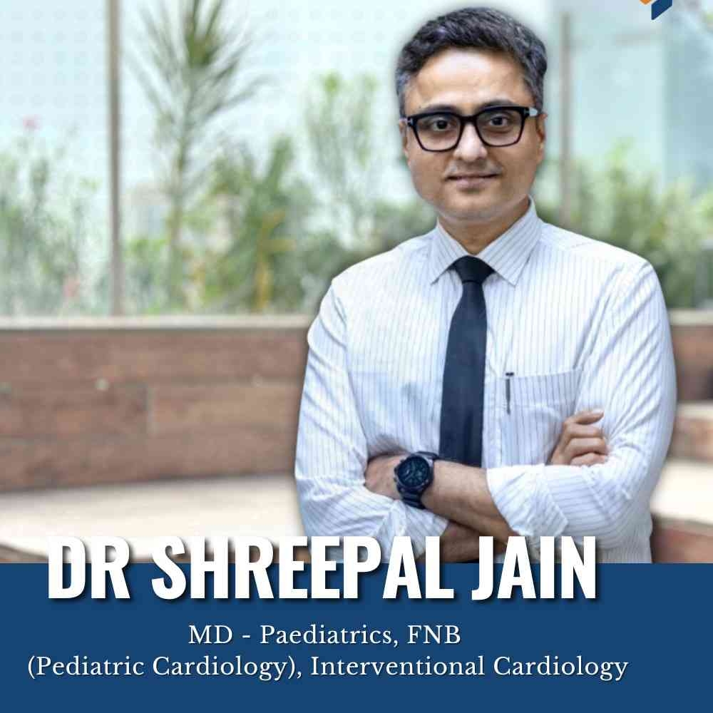
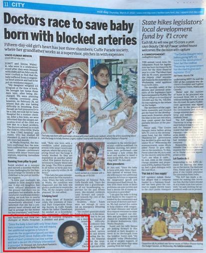
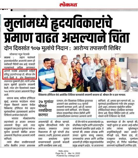
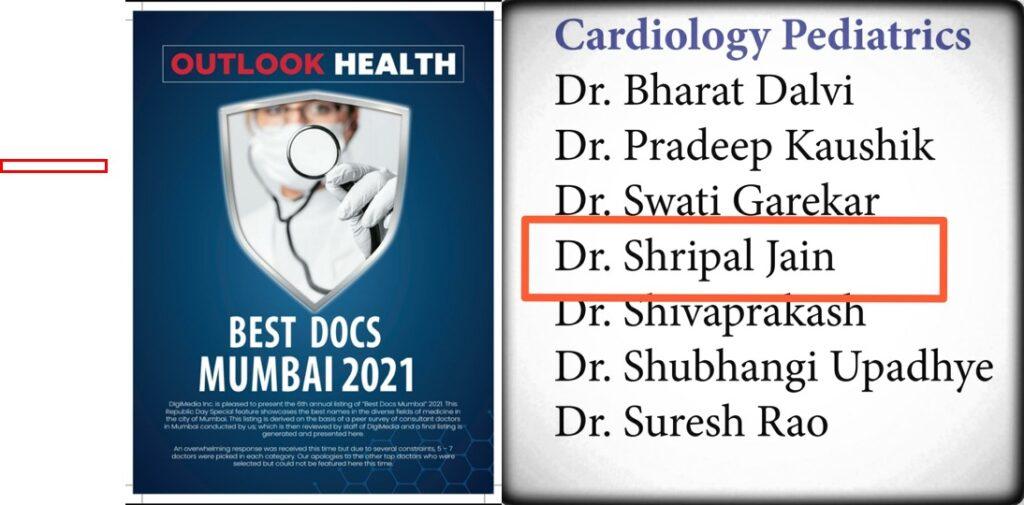
Dr. Shreepal Jain

Dr. Shreepal Jain

PEDIATRIC CARDIOLOGY

Phone +91-9326171030

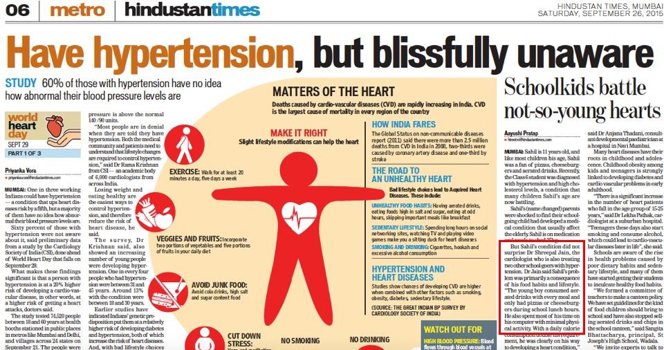
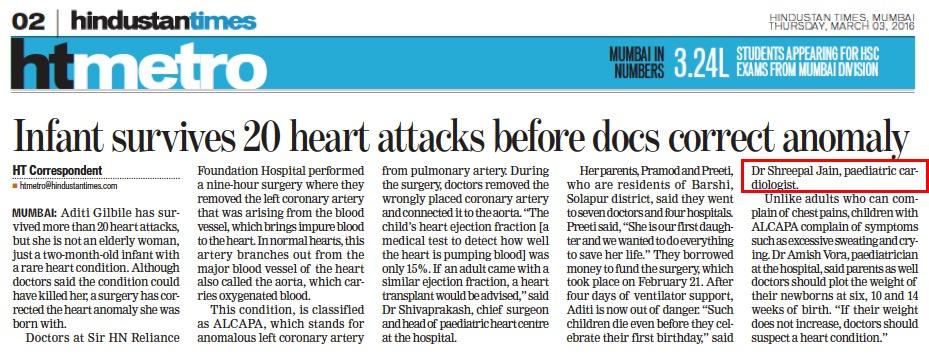
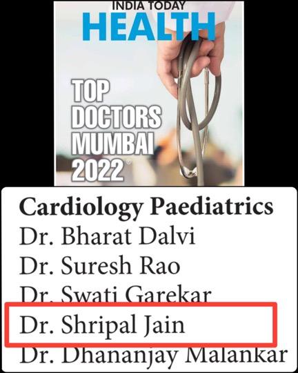
Dr. Shreepal Jain is a renowned Paediatric Interventional and Fetal Cardiologist practicing in Mumbai since last 16 years. Dr. Jain has received several distinctions during his undergraduate training. He had topped his batch during second and third year of MBBS. He was awarded the P. N. Khanna Memorial award for securing highest marks in Forensic Medicine. After completing his MBBS, Dr. Jain was trained in Paediatrics at the Topiwala National Medical College and BYL Nair Hospital, Mumbai. Later he pursued his formal Paediatric Cardiology training from the prestigious Amrita Institute of Medical Sciences, Kochi after securing All India First rank in the entrance test of the National Board of Examination. He received further training in Interventional Paediatric Cardiology and Pulmonary Hypertension from Stollery Children’s Hospital, Edmonton, Canada. During his tenure in Canada, he was awarded the SCAI (Society of Cardiac angiography and Interventions) scholarship for fellows training in Las Vegas. He has presented multiple papers at National and International Conferences.

16, 1st Floor, Tender Hearts Clinic, Avighna IX, Opposite Ladoo Samrat Restaurant, Dr Baba Saheb Ambedkar Rd, Lal Baug, Mumbai, Maharashtra, India

Testimonials

Dr.Shripal Jain Sir I have seen….A wonderful personalityI am always inspired by the love and devotion towards one's work and the radiance on his face.Sir saved my son's lifeSir for me*Heartbroken Narayan*.. Omniscient.

A wonderful personality

Good experienced doctor and very kind person,Explain very clearly everything in depth.

Good Experienced Doctor

Latest Update

Congenital Heart Defect Treatment in Parel – Early Diagnosis Matters

It's Done!!

Your appointment is confirmed

Doctor Name:

Date & Time:

Contact:

Address: 16, 1st Floor, Tender Hearts Clinic, Avighna IX, Opposite Ladoo Samrat Restaurant, Dr Baba Saheb Ambedkar Rd, Lal Baug, Mumbai, Maharashtra, India

Appointment details has also been shared with you on your mobile number . Please arrive atleast ten minnutes ahead of the scheduled time.

treatments